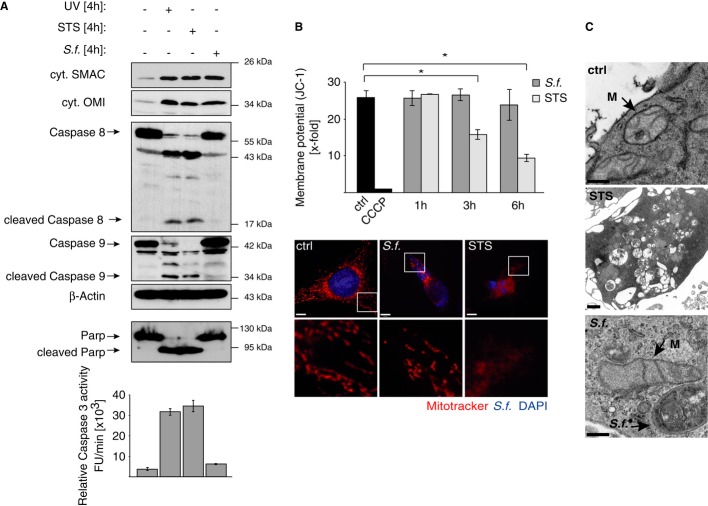
Figure 3. Intracellular Shigella induces the release of mitochondrial SMAC without inducing mitochondrial damage.
- HeLa wt cells were left untreated (−), treated with UV light (10 mJ/m2), STS (0.5 μM) or were infected with Shigella M90T (MOI 30). After 4 h SMAC, OMI, Caspase-8 and Caspase-9 were analyzed in the cytosolic fractions by Western blotting. Actin served as a loading control. PARP cleavage was analyzed in nuclear fractions by Western blotting. Caspase-3 activity was determined fluorimetrically in cytosolic fractions using the substrate Ac-DEVD-AFC. Data are presented as mean ± SEM (n = 3).
- HeLa wt cells were left untreated (ctrl), infected with Shigella M90T (MOI 30) or treated with STS (0.5 μM). ΔΨm was analyzed after JC-1 or MitoTracker staining at the indicated time points using FACS (upper panel) or confocal microscopy (lower panel), respectively after 6 h. Control cells were treated with CCCP (50 mM). In confocal images, Shigella was stained blue by immunofluorescence. Data are presented as mean ± SEM (n = 3); *P < 0.05; (scale bar = 8 μm)
- HeLa wt cells were left untreated (ctrl), treated with STS (0.5 μM) or were infected with Shigella M90T (MOI 30). Mitochondrial cristae structure was analyzed 6 h p.i. by transmission electron microscopy. Arrows indicate mitochondria (M) or Shigella (scale bar: 300 nm (ctrl, S.f.); 1 μm (STS)).
Source data are available online for this figure.

