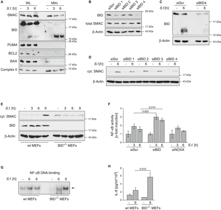
Figure 4. BID induces mitochondrial release of SMAC in Shigella-infected cells.
- HeLa wt cells were left untreated (−) or were infected with Shigella M90T (MOI 30). At the indicated time points whole-cell lysates (WL) and isolated mitochondria (Mito) were analyzed by Western blotting.
- HeLa wt cells were transiently transfected with four different siRNAs against BID (siBID1–4) or non-targeting ctrl (siScr). After 48 h cells were analyzed by Western blotting.
- HeLa wt cells were transiently transfected with BID-siRNA4. After 48 h cells were left untreated (−) or were infected with Shigella M90T (MOI 30). The mitochondrial fraction was analyzed 6 h p.i. by Western blotting.
- HeLa wt cells were transiently transfected with four different siRNAs against BID (siBID1–4) or non-targeting ctrl (siScr). After 48 h cells were left untreated (−) or were infected with Shigella M90T (MOI 30). Cytosolic extracts were analyzed 6 h p.i. by Western blotting.
- MEFs isolated from wt or BID−/− mice were left untreated (−) or infected with Shigella M90T (MOI 50). Cytosolic fractions were analyzed by Western blotting at the indicated time points p.i..
- HeLa wt cells were transiently transfected with specific siRNAs for BID, NOXA or non-targeting ctrl (siScr). After 48 h cells were infected with Shigella M90T (MOI 30). NF-κB DNA binding activity was analyzed by ELISA at the indicated time points p.i.. Data are presented as mean ± SD (n = 3).
- MEFs were treated as in (E). NF-κB DNA binding activity was analyzed by EMSA at the indicated time points p.i..
- MEFs were treated as in (E). IL-6 secretion was monitored by ELISA in supernatants of the cells 9 h p.i..
Source data are available online for this figure.

