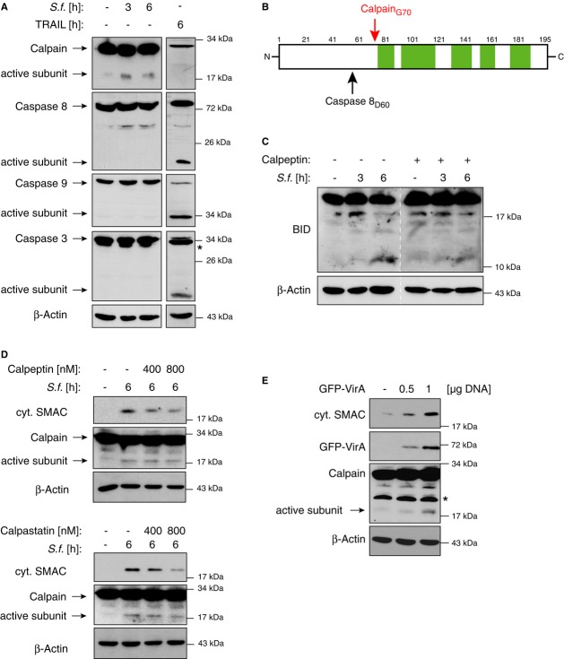
Figure 5. BID is cleaved by Calpain after infection with Shigella.
- HeLa wt cells were left untreated (−), infected with Shigella M90T (MOI 30) or stimulated with TRAIL (50 ng/ml). At the indicated time points cytosolic extracts were analyzed by Western blotting (* marks unspecific bands).
- Schematic sequence coverage (green areas) of BID peptides identified by mass spectrometry. Caspase-8 and Calpain cleavage sites are indicated.
- HeLa wt cells were infected with Shigella M90T (MOI 30) and were exposed to the calpain inhibitor calpeptin (400 nM) after 30 min (time point zero, see Materials and Methods). Mitochondrial fractions were analyzed by Western blotting at the indicated time points p.i..
- HeLa wt cells were left untreated (−) or were infected with Shigella M90T (MOI 30) in combination with calpeptin (upper panel) or calpastatin (lower panel) at the indicated concentrations. Cytosolic extracts were analyzed by Western blotting 6 h p.i..
- HeLa wt cells were mock transfected (−) or transiently transfected with the indicated amounts of GFP-VirA. After 40 h cytosolic fractions were analyzed by Western blotting (* marks unspecific bands).
Source data are available online for this figure.

