
Figure 6.
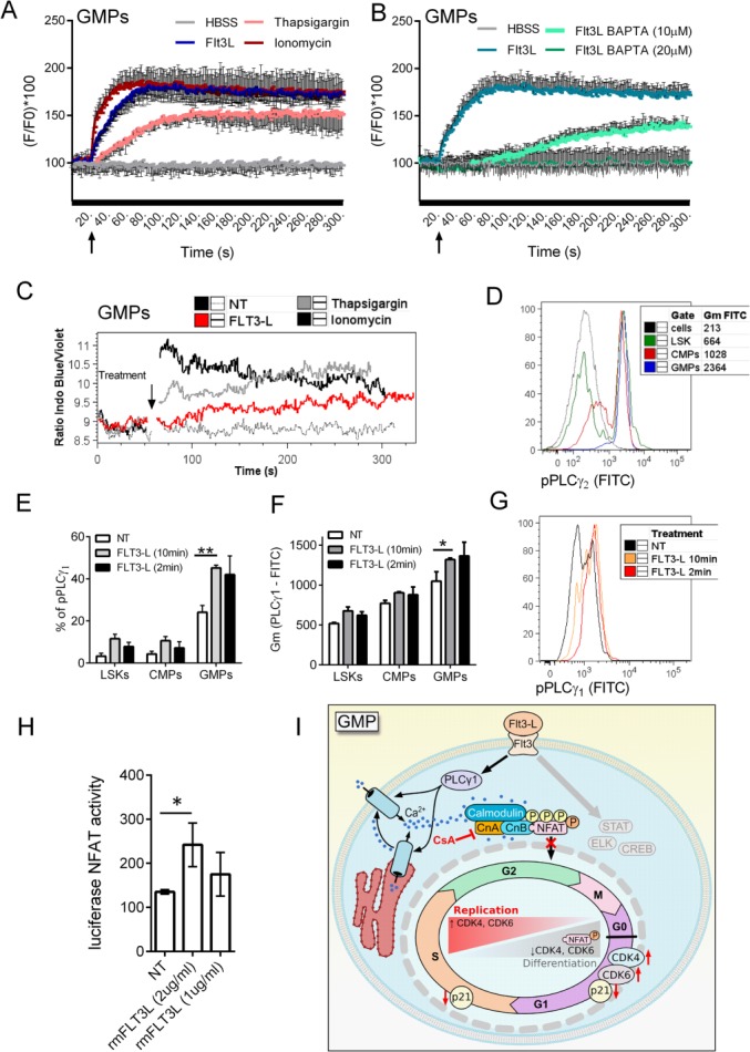
Flt3-L triggers signaling leading to nuclear factor of activated T cells (NFAT) translocation in granulocyte–monocyte progenitors (GMPs). (A): Levels of intracellular Ca2+ measured as Fluo4 fluorescence in sorted lin−Sca1+cKIT+ bone marrow (BM) cells (LSKs), common myeloid progenitors (CMPs), and GMPs. After 20 seconds of measurements cells were triggered with FLT3-L, ionomycin, thapsigargin, or Hanks' balanced salt solution (HBSS), and Ca2+ levels were assessed for another 5 minutes. (B): Intracellular Ca2+ chelator BAPTA was used to block Ca2+ release induced by FLT3-L in sorted GMPs. (C): Flt3-L induces Ca2+ release in GMPs sorted as lin−, cKit+, Sca-1−, CD34+, CD16/32high. Graph of Ca2+ release analysis in GMPs from lineage-depleted BM cells. Flt3-L (1μg/ml) was added after 1 minute of measurement followed by 4 minutes Ca2+ release measurement. Representative of three independent experiments, where BM cells from five mice were pooled. (D): Different levels of phospholipase Cγ (PLCγ2) phosphorylation (pPLCγ2) in LSKs (pool of hematopoietic stem cell [HSCs] and multipotent progenitors [MPPs]; lin−, cKit+, Sca-1+), CMPs, and GMPs in freshly isolated BM. (E–G): Different levels of pPLCγ1 in progenitor populations before and after Flt3-L administration. Lineage-depleted BM cells were cultured for 4 hours in HSC medium, Flt3-L (1 μg/ml) was added 15 minutes before fixing and labeling with antibodies for progenitor markers, PLCγ1 and pPLCγ2. Cells were gated as LSKs (pool of HSCs and MPPs; lin−, cKit+, Sca-1+), CMPs (lin−, cKit+, Sca-1−, CD34+, CD16/32int), and GMPs (lin−, cKit+, Sca-1−, CD34+, CD16/32high). (E, F): Percentage of pPLCγ1 cells and Gm of fluorescence of pPLCγ1 and double labeling of total PLCγ1 with pPLCγ1 upon trigger with Flt3-L are shown. Data are presented as mean ± SE, *, p < .05 and **, p < .01 in an unpaired Student's t-test. (G): Histogram overlay of pPLCγ1 intensity in GMPs after the Flt3-L trigger. Representative experiment from three independent experiments is shown. (H): Flt3-L induces NFAT translocation in cKIT+-enriched BM cells. BM cells were depleted of lineaged cells and enriched with cKIT+ beads, maintained in HSC medium and transduced with NFAT reporter constructs. Transfected cells were kept in HSC medium for 48 hours, and 1 μg/ml of Flt3-L was added for last 4 hours of culture before nuclear translocation of NFAT reflecting activity of luciferase reporter gene was measured. Data are presented as mean ± SE from one representative of three independent experiments, *, p < .05 in an unpaired Student's t-test. BM pooled from 10 mice was used for each experiment. (I): Proposed graphical scheme of cell cycle regulation in GMPs. Flt3-L activates PLCγ1 and induce increase in intracellular Ca2+ levels, this further activates calcineurin and NFAT translocation. When calcineurin-NFAT interaction is blocked, expression of cell cycle regulation genes in GMPs is changed to promote further proliferation. Abbreviations: CMP, common myeloid progenitors; CsA, Cyclosporine A; FITC, fluorescein isothiocyanate; GMP, granulocyte–monocyte progenitor; HBSS, Hanks' balanced salt solution; LSK, lin−Sca1+cKIT+ bone marrow cells; NFAT, nuclear factor of activated T cells; NT, non-treated; PLC, phospholipase Cγ.