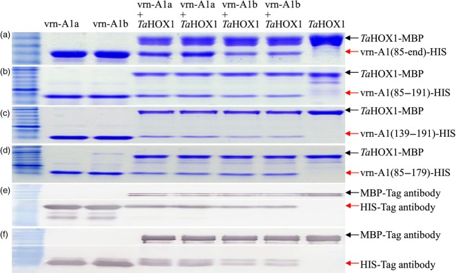
Figure 4.

In vitro interaction between TaVRN-A1 and TaHOX1 proteins.(a–d) Pull-down assays of TaHOX1-MBP and TaVRN-A1 interactions. In vitro interaction between TaHOX1(100–end) and TaVRN-A1(85–end) (a), TaVRN-A1(85–191) (b), TaVRN-A1(139–191) (c), and TaVRN-A1(85–179) (d).(e, f) Protein immunoprecipitation analyses of TaHOX1 and TaVRN-A1. TaHOX1-MBP was detected by anti-MBP antibody (the upper panel, e and f). TaVRN1-HIS(85–end) and TaVRN-A1-HIS(139–191) were detected by anti-HIS antibody (the lower panel, e and f). Lane 1, purified Jagger vrn-A1a-HIS-tag; lane 2, purified 2174 vrn-A1b-HIS-tag; lanes 3 and 4, interaction of TaVRN-A1a-HIS-tag and TaHOX1-MBP-tag; lanes 5 and 6, interaction of TaVRN-A1b-HIS-tag and TaHOX1-MBP-tag; lane 7, purified TaHOX1-MBP-tag; M, protein marker. Arrowheads represent expressed or interacting proteins. Six independent replicates were performed for each interaction, and two replicates are shown to indicate consistency among replicates.
