
Figure 5.
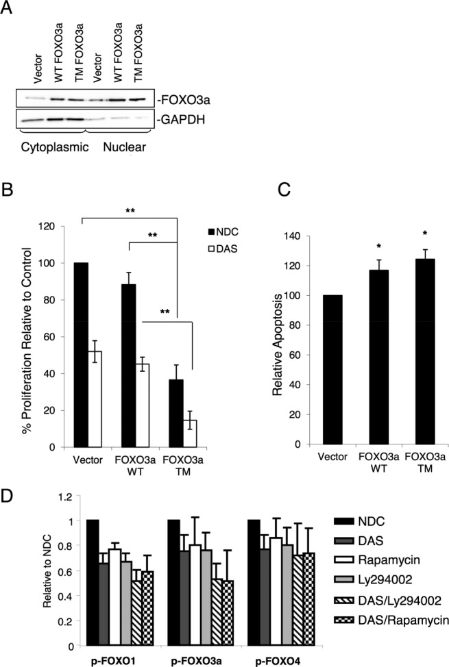
Over-expression of FOXO3a induces strong inhibition of proliferation in the K562 cell line. (A): FOXO3a WT, FOXO3a TM (a constitutively active mutant), and vector control were transiently over-expressed in K562 cells and cytoplasmic versus nuclear fractionation carried out. Following isolation, 10 µg of nuclear and cytoplasmic lysates were separated by SDS PAGE and the distribution of FOXO3a determined by Western blotting. GAPDH was used as a cytoplasmic marker. (B): Cells transfected with FOXO3a WT or TM were treated or not with dasatinib (DAS, 10 nM) for 24 hours and BrdU used to assess proliferation (n = 3, **, p < .01). (C): Levels of apoptosis were determined by Annexin V/7-AAD staining (n = 3, *, p ≤ .05). (D): CML CD34+ cells were treated for 24 hours with rapamycin (10 nM), LY294002 (25 µM), DAS (150 nM), or the combination of DAS with either LY294002 or rapamycin, and FOXO1, 3a, and 4 phosphorylation were measured by flow cytometry (n = 3). Statistical analysis for each treatment is reported in Supporting Information Figure S2. Abbreviations: DAS, dasatinib; NDC, no drug control.