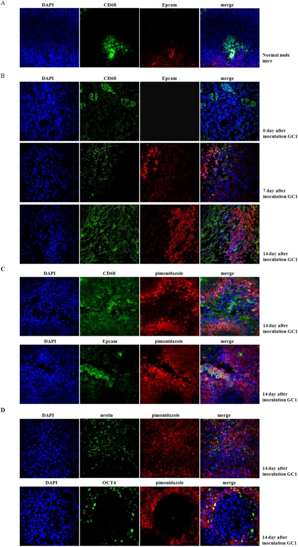
Figure 4.

Peritoneal milky spots (PMSs) served as a hypoxic niche during gastric cancer peritoneal dissemination (GCPD). (A): Normal mouse peritoneum was fixed and sectioned. CD68 (marked PMS macrophage) and pimonidazole incorporation (marked hypoxia region) were analyzed by immunofluorescence staining (DAPI showed blue staining, CD68 showed green staining, and pimonidazole showed red staining, magnification ×400). (B): Fifteen male BALB/c nude mice (8–10 weeks old) were intraperitoneally (i.p.) injected with 1 × 10⁴ GC1sps to establish a GCPD time-dependent model. Peritoneal tumor-bearing mice were sacrificed on days 7, 14, and 21 (five mice at each time point). Two hours before sacrifice, 60 mg/kg pimonidazole hydrochloride was injected intravenously (i.v.) into tumor-bearing mice to detect the hypoxic region. Mouse peritoneum was fixed and sectioned. CD68 and Epcam (marked GCSPC) expression were analyzed by immunofluorescence staining to mark macrophage and GCSPC, respectively (DAPI showed blue staining, CD68 showed green staining, and pimonidazole showed red staining, magnification ×400). (C): CD68, Epcam expression, and pimonidazole incorporation were analyzed by immunofluorescence staining (DAPI showed blue staining, CD68 and Epcam showed green staining, and pimonidazole showed red staining, magnification ×600). (D): Stem-related protein OCT4 and Nestin expression and pimonidazole incorporation were analyzed by immunofluorescence staining (DAPI showed blue staining, OCT4 and Nestin showed green staining, and pimonidazole showed red staining, magnification ×600).
