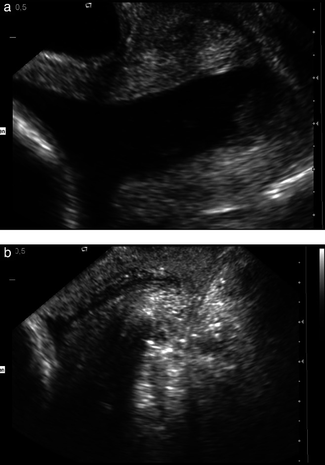
Figure 3.

Transvaginal sonography of a cervix with U-shaped complete funneling and sludge in a nulliparous patient at 24 weeks' gestation before (a) and after (b) pessary placement (proximal inner diameter 35 mm, height 21 mm, distal outer diameter 65 mm), showing closer attachment, which suggests normal cervical gland area after placement of pessary. The patient delivered at 37 weeks after pessary removal.
