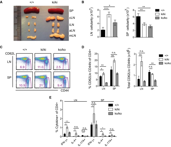
Figure 5. Mice expressing catalytically inactive Malt1 have enlarged lymph nodes and accumulate T cells with an activated phenotype.
- A, B Analysis of the anatomical size (A) and cellularity (B) of lymph nodes (LN; c: superficial cervical, i: inguinal, a: axillary, m: mesenteric) and spleens (SP) of wild-type (+/+, n = 4) and Malt1 knock-in (ki/ki, n = 4) mice at the age of 8 (A) and 6 (B) weeks.
- C Analysis of the expression of CD62L and CD44 on CD4+ cells isolated from lymph nodes (LN) or spleens (SP) of 6-week-old mice of the indicated genotypes.
- D Quantification of data shown in (C), obtained with wild-type (+/+, n = 4), Malt1 knock-in (ki/ki, n = 4), and knock-out (ko/ko, n = 3) mice.
- E Intracellular cytokine expression in CD4+ cells isolated from the lymph nodes (LN) or spleens (SP) of 6-week-old mice of wild-type (+/+, n = 4), Malt1 knock-in (ki/ki, n = 4), and knock-out (ko/ko, n = 3) mice.
Data information: Bars represent means ± SD; *P < 0.05; **P < 0.005; ***P < 0.0005; n.s., not significant (unpaired t-test). Data are representative of six (A, B), four (C, D), and two (E) experiments.

