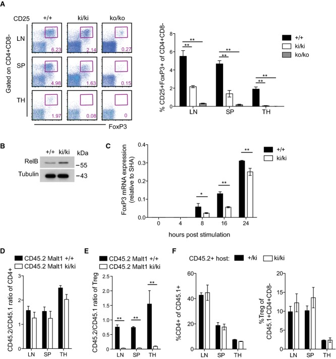
Figure 7. Malt1 C472A knock-in mice have a cell-intrinsic defect in Treg development.
- A Analysis of wild-type (+/+, n = 4), knock-in (ki/ki, n = 4), and knock-out (ko/ko, n = 3) mice for the presence of thymic (TH) and peripheral CD4+CD25+Fop3+ Treg cells. SP, spleen; LN, lymph node.
- B Western blot analysis of RelB expression in total thymocytes of wild-type and knock-in mice.
- C FoxP3 mRNA levels in naïve CD4+ peripheral T cells after stimulation with anti-CD3 and anti-CD28 in the presence of TGF-β and IL-2.
- D, E Analysis of CD45.1 wild-type mice that were lethally irradiated and reconstituted with a 1:1 ratio of wild-type CD45.1+ to CD45.2+ wild-type (+/+, n = 3) or knock-in (ki/ki, n = 4) bone marrow cells, 8 weeks after transfer. Cells from lymph nodes (LN), spleen (SP), and thymus (TH) were analyzed. The CD45.1/CD45.2 ratio was determined in total CD4+ T cells (D) and in CD4+CD25+FoxP3+ Treg cells (E).
- F CD45.2 control (+/ki) or Malt1 knock-in mice (ki/ki) were lethally irradiated and reconstituted with wild-type CD45.1+ bone marrow cells (reciprocal chimeras) and T-cell development analyzed 8 weeks after transfer (n = 3).
Data information: Bars represent means ± SD; *P < 0.05; **P < 0.005; n.s., not significant (unpaired t-test). Results are representative of four (A), two (B, C, F), and three (D, E) experiments.
Source data are available online for this figure.

