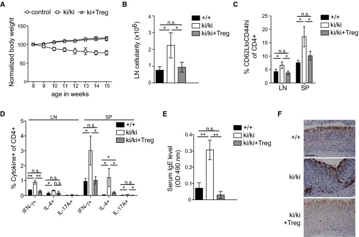
Figure 8. Treg transfer rescues autoimmune symptoms in Malt1 C472A knock-in mice.
- A Weight curves of untreated wild-type and heterozygous mice (control, n = 3 for each) and of knock-in mice (ki/ki) with (n = 3) or without (n = 4) transfer of 106 Treg cells at the age of 8 days.
- B–E Analysis of the total lymph node (LN) cellularity (per gram body weight) (B), the percentage of CD4+ cells with a CD62LloCD44hi phenotype (C), the percentage of cytokine-positive CD4+ cells isolated from lymph nodes (LN) or spleens (SP) (D) and the serum IgE levels (E) of 5-week-old wild-type (+/+) and knock-in (ki/ki) mice, and of knock-in mice having received an adoptive transfer of 106 Treg cells (ki/ki + Treg) at the age of 3 days (n = 3).
- F Immunohistochemical analysis of CD3+ cells in stomach sections of mice described in (B–E). Scale bar, 100 μm.
Data information: Bars represent means ± SD; *P < 0.05; **P < 0.005; n.s., not significant (unpaired t-test). Results are representative of two experiments (A-F).

