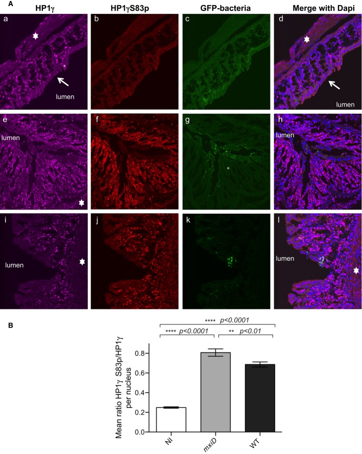
Figure 1. HP1γ immunostaining in the distal colon of guinea pigs following intra-rectal challenge with Shigella flexneri strains.
- Samples of the distal colon were taken 7 h after infection with the T3SS defective mxiD Shigella pGFP(e–h), WT Shigella pGFP (i–l) or PBS treated as control (a–d) and co-stained with anti-HP1γ (a, e, and i) or anti-HP1γS83p (b, f, and j) antibodies and merged with DAPI (d, h, and l). GFP allowed for visualization of the bacterium (c, g, and k). The star indicated the submucosa, and the arrow showed the terminally differentiated columnar absorptive enterocytes devoid of HP1γ.
- Fluorescence intensity ratios between HP1γS83p and total HP1γ. HP1γS83p staining was quantified on HP1γ-positive nuclei in each field. The intensity of fluorescence for each channel of interest was measured, and the ratio of fluorescence intensity between HP1γS83p and total HP1γ signals was calculated as described in Materials and Methods. Statistical analysis was performed as described in Materials and Methods.

