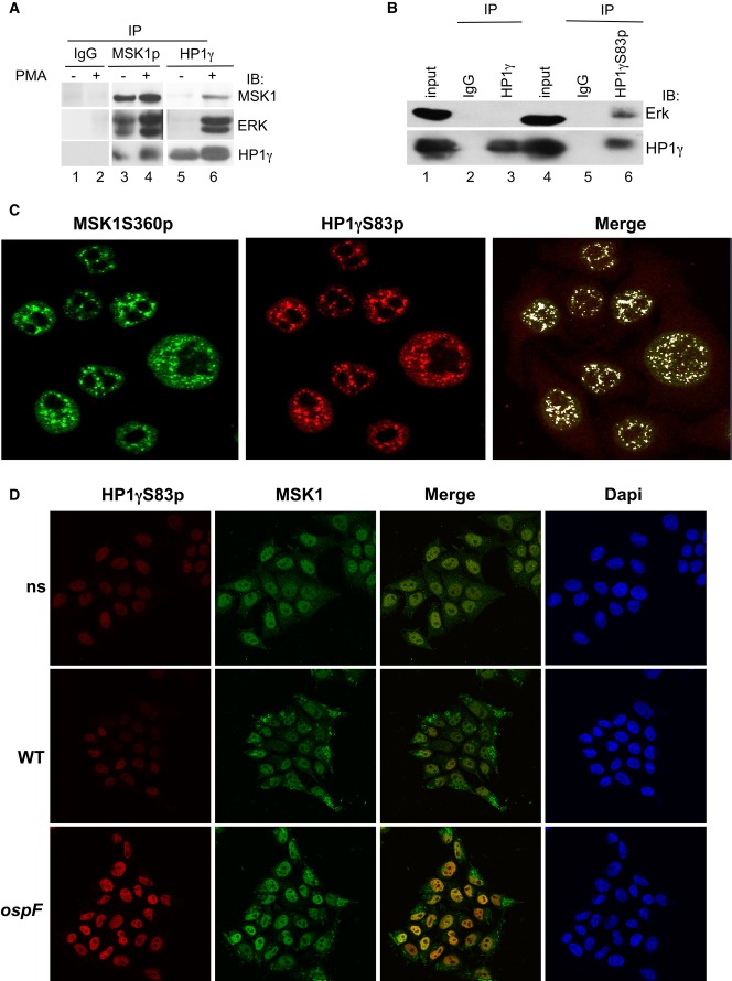
Figure 5. MSK1 forms a molecular complex with the phosphorylated pool of HP1γ in cells.
- PMA stimulation leads to the formation of molecular tri-complex between the HP1γ and the ERK/MSK1 kinases. HeLa cells were stimulation by PMA (60 min), and cellular extracts were immunoprecipitated with mouse IgG as a negative control or anti-MSK1S360p or anti-HP1γ antibodies. Western blots were performed with anti-MSK1, anti-ERK, and anti-HP1γ antibodies.
- The phosphorylated pool of HP1γ pulls down the ERK kinase. Cellular extracts from HeLa cells were immunoprecipitated with mouse IgG as a negative control or anti-HP1γS83p, or anti-HP1γ antibodies. Western blots were performed with anti-ERK and anti-HP1γS83p antibodies.
- Confocal microscopy showing high coincidence of immunostaining between HP1γS83p and active MSK1. Immunofluorescence was performed with anti-HP1γS83p (red) and anti-MSK1S360p (green) antibodies.
- Confocal microscopy showing the immunostaining between HP1γS83p (red) and MSK1 upon infection with the WT and ospF mutant strains (green).
Source data are available online for this figure.

