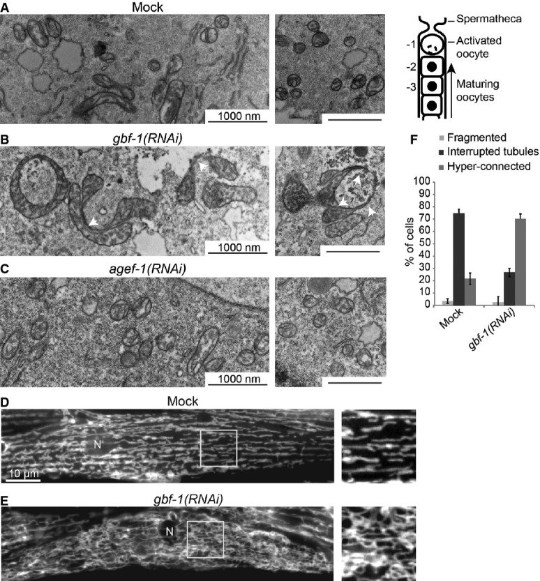
Figure 1. GBF-1 affects mitochondrial morphology.

A–C Electron microscopy (EM) of mitochondria in Caenorhabditis elegans oocytes. Wild-type is shown in (A). On the right, a schematic view of the C. elegans proximal gonad is shown. Oocytes indicated with -2 were used for EM analysis. The depletion of GBF-1 (B) leads to the formation of enlarged mitochondria connected through thin membrane connections (indicated with white arrowheads). The mitochondria of agef-1(RNAi) worms (C) appear unaffected.
D, E Live imaging of TOM70::GFP in C. elegans muscle cells. In wild-type muscle cells (D), the mitochondrial network is organized in interrupted slightly branched tubules. gbf-1(RNAi) caused disorganization of the mitochondrial network and an increase of connections between mitochondria (E). N, nucleus.
F Quantification of (D) and (E) of three independent experiments, showing the percentage of muscle cells in a total of N = 392 (Mock) and N = 466 [gbf-1(RNAi)] cells, with fragmented, tubular or hyper-connected mitochondria. Error bars represent standard deviation.
