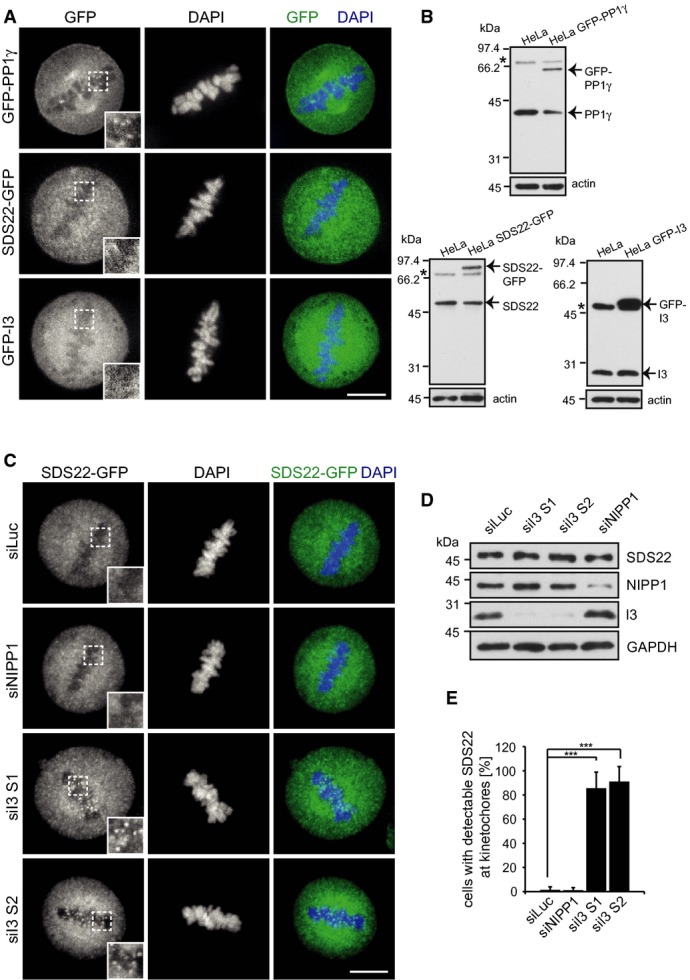
Figure 4. Depletion of I3 induces re-localization of SDS22 to kinetochores.

- Neither SDS22 nor I3 colocalize with PP1γ at metaphase kinetochores under control conditions. Stable HeLa cell lines expressing GFP fusions of indicated proteins were fixed and DAPI-stained. SDS22-GFP is expressed under its own promoter in BAC transgenic cells. For localization images of GFP-I3-expressing cells in all mitotic stages see also Supplementary Fig S2. Representative confocal images. Scale bar, 10 μm.
- Expression levels of GFP fusions relative to endogenous proteins in Western blots stained with specific antibodies. Actin served as a loading control. Asterisks denote non-specific bands.
- SDS22 re-localization upon I3 depletion. SDS22-GFP cells were treated with indicated siRNAs and processed as in (A). Representative images. Note the SDS22-GFP signal at kinetochores in I3, but not control-depleted cells. Scale bar, 10 μm.
- Depletion efficacy for individual siRNAs as determined by Western blot.
- Quantification of (C). Error bars indicate s.d. of three independent experiments with 25 cells per condition. ***P < 0.001.
Source data are available online for this figure.
