
Fig. 2.
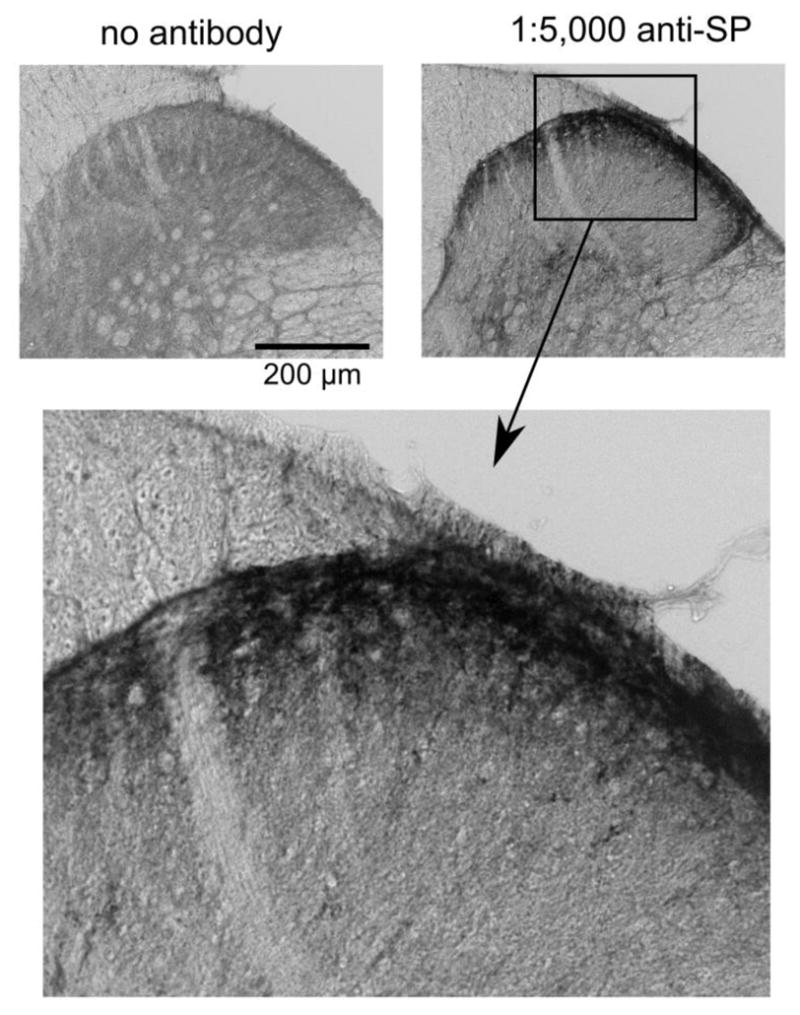
Representative substance P (SP) immunolabeling in the substantia gelatinosa of the musk shrew spinal cord, the terminal location of spinal afferent fibers [46]. The images show the effects of removing the primary antibody from the immunohistochemistry protocol compared to including a concentration of 1:5,000. Lower image is a higher magnification of the SP labeling in the upper right panel.