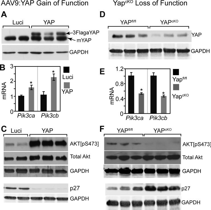
Figure 3. YAP is sufficient and required for activating the PI3K-AKT pathway.
A. AAV9:YAP-mediated overexpression of activated YAP. AAV9:YAP was subcutaneously injected into P3 pups. 7 days later, hearts were collected for western blotting. AAV9:Luci served as a negative control. B. qRTPCR measurement of Pik3cb and Pik3ca mRNA level in hearts of mice treated with AAV9:YAP or AAV9:Luci. *, P<0.05. n=4. C. Western blot assessment of AKT pathway activation state in hearts of AAV9:YAP or AAV9:luci-treated mice. Primary antibodies were directed against AKT[pS473] (activated AKT), total AKT, GAPDH (internal loading control), or p27. D. YAP protein level in hearts from 4-week-old YapcKO mice and their littermate controls (Yapfl/fl). E. qRT-PCR measurement of Pik3cb and Pik3ca mRNA level in YapcKO and Yapfl/fl heart. *, P<0.05, n=3. F. Western blot assessment of AKT pathway activation state in YapcKO and Yapfl/fl heart.

