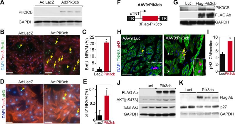
Figure 4. Pik3cb overexpression increased cardiomyocyte proliferation.
A. Adenovirus-mediated Pik3cb overexpression in cultured NRVMs isolated on postnatal day 4 (P4). PIK3CB protein was detected by immunoblotting. B-E. Effect of Pik3cb overexpression on P4 NRVM proliferation in vitro was measured by BrdU incorporation rate (B, C) and phosphohistone H3 positive (pH3+) cardiomyocyte frequency (D,E). Bar = 50 μm. Arrows indicate BrdU and pH3 positive cardiomyocytes. Arrowheads indicate non-myocytes. *, P<0.05. n=3. F. Schematic of the AAV9:Pik3cb construct. 3Flag epitope-tagged Pik3cb was expressed from the cardiac troponin T promoter. G. AAV9:Luci and AAV9:Pik3cb were injected subcutaneously into P2 neonatal mice. 8 days later, hearts extract western blots were probed with FLAG, PIK3CB, or GAPDH antibodies. H, I. Effect of Pik3cb overexpression on neonatal cardiomyocyte proliferation in vivo. AAV9:Luci or AAV9:Pik3cb were administered subcutaneously to P2 neonatal mice. Hearts were analyzed by immunofluorescent staining at P9. Arrows and arrowheads indicate pH3+ cardiomyocytes and non-myocytes, respectively. Boxed area is enlarged in inset. Bar = 50 μm. *, P<0.05. n=4. J,K. Immunoblot analysis of P9 heart lysates. Mice were treated with AAV:Luci or AAV9:Pik3cb at P2.

