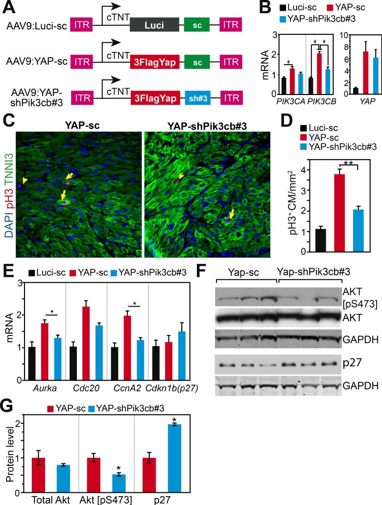
Figure 6. Yap stimulation of mouse cardiomyocyte proliferation requires Pik3cb in vivo.
A. Schematic view of AAV plasmids. shRNA effective for Pik3cb knockdown (shPik3cb#3) was cloned into AAV ITR plasmid downstream of YAP[S127A] (human) to simultaneously express YAP and knockdown Pik3cb. Control constructs contained Luci (luciferase) or scrambled control shRNA (sc). B. qRT-PCR measurement of Pik3cb, Pik3ca and Yap mRNA levels from P9 mouse heart. AAV was injected into mice at P2. Primers that amplified both mouse and human Yap were used for measuring Yap expression level. C, D. pH3 immunofluorescence staining of heart sections. AAV was injected into 2-dayold mouse pups and hearts were analyzed at P9. E. qRT-PCR measurement of cell cycle genes from P9 hearts after AAV treatment at P2. F, G. Effect of Pik3cb knockdown in the presence of activated YAP on activated Akt and p27 protein levels. B, D, E, n=4. *, P<0.05.

