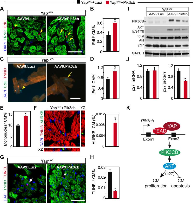
Figure 8. AAV9:Pik3cb increased cardiomyocyte proliferation and decreased cardiomyocyte apoptosis in the absence of YAP.
YapcKO mice were treated with AAV9:Pik3cb or AAV9:Luci as indicated in Fig. 7A. A-F. Effect of Pik3cb overexpression on depressed cardiomyocyte proliferation seen in YapcKO heart. Cardiomyocyte proliferation was measured by uptake of EdU (A-D) 4 weeks after birth. A-B. EdU staining and quantification. EdU was administered once at P21. C-D. EdU quantification with dissociated cardiomyocytes. EdU was administered at P21 and P24. Arrows and arrowheads indicate EdU positive cardiomyocytes and non-myocytes, respectively. E. Percentage of mononuclear cardiomyocytes. F. Measurement of cytokinesis. Aurora kinaseB antibody was used to detect cardiomyocytes undergoing cytokinesis. G-H. Effect of Pik3cb overexpression on cardiomyocyte apoptosis in YapcKO heart, as measured by TUNEL assay. Arrows and arrowheads indicate TUNEL+ cardiomyocytes and nonmyocytes, respectively. I. Effect of Pik3cb overexpression on AKT activation and p27 expression in YapcKO heart. Lysates collected from one-month old mouse hearts were analyzed by western blotting. J. Effect of Pik3cb overexpression on p27 mRNA and protein levels in YapcKO heart. K. YAP-TEAD stimulated Pik3cb transcription through an enhancer in the first intron. Pik3cb activates AKT, which suppresses cardiomyocyte proliferation and increases cardiomyocyte apoptosis, in part through increased p27 protein. Bar = 50 μm. *, P<0.05. n=3.

