
Figure 2. TDP-1 limits the amount of nuclear dsRNA.
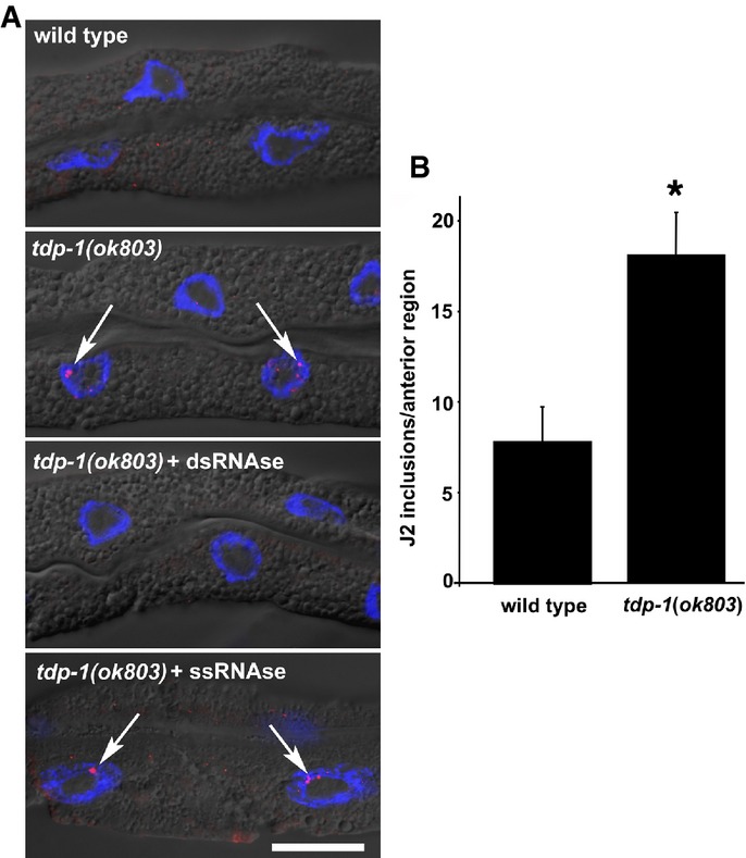
- Fixed, isolated intestinal tissue probed with anti-dsRNA antibody (J2). The J2 antibody recognizes dsRNA stretches of 40 bp or more in a sequence-independent manner. Intensely stained inclusions (red dots, indicated by arrows) were detected in intestinal nuclei (blue, DAPI counterstain) specifically in tdp-1(ok803) mutant worms (middle panel). J2-reactive inclusions were observed in 28% tdp-1(ok803) intestinal nuclei scored (30/107), but not detected in wild-type controls (0/122) or tdp-1(ok803) (0/105) fixed tissue pretreated with dsRNA nuclease (V1) before J2 staining. J2-reactive foci were still observed (arrows, bottom panel) in intestinal nuclei pretreated with the ssRNA-specific nuclease (T1) (29%, 12/41). Scale bar, 20 μm.
- Quantification of anterior J2 inclusions using ImageJ software. tdp-1(ok803) worms had significantly more J2 inclusions (*P < 0.01, Student's t-test, error bars = SEM). Representative projection images used to generate this data are shown in Supplementary Fig S4.