
Fig. 5.
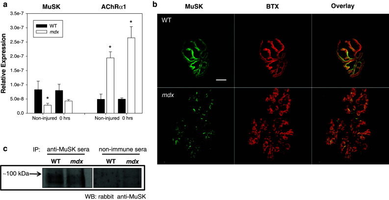
Presence of muscle specific kinase (MuSK) in healthy (WT) and dystrophic (mdx) muscle. a Transcripts for acetylcholine receptors (AChRs) and muscle specific kinase (MuSK) were quantified using qRT-PCR. Relative expression of MuSK was decreased in mdx quadriceps muscles when compared to WT. MuSK, which is important for proper clustering of acetylcholine receptors, may also contribute to the abnormal NMJ morphology seen in mdx mice. Interestingly, AChR transcripts were also increased. Expression was unchanged for both AChRs and MuSK following injury (see supplemental for later time points). All data are presented as mean ±SD, p < 0.05. * indicates statistical significance from WT. b Uninjured neuromuscular junctions (NMJs) were stained with α-Bungarotoxin (BTX, red) and labeled with antibodies against muscle specific kinase (MuSK, green). NMJs in mdx mice show a decrease in MuSK labeling, compared with NMJs from WT mice. Scale bar equals 10 µm. c MuSK was immunoprecipitated from extracts of uninjured quadriceps muscle from WT or dystrophic mice, then immunoblotted with anti-MuSK antibodies. Ctrl: immunoprecipitate from muscle probed with a non-immune serum. Equal loading and transfer was confirmed by Ponceau S staining (not shown)