Fig. 8.
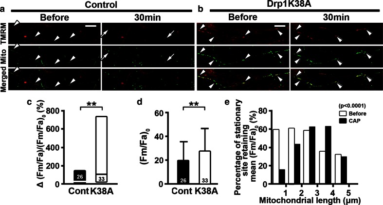
Mutant Drp1K38A overexpression retains mitochondrial membrane potential upon capsaicin treatment. a, b Time-lapse images of axonal mitochondria labeled with Mito-Dendra2 (Mito) and stained with TMRM for mitochondrial membrane potential. TMRM labeling co-localized with axonal mitochondria (arrowheads). While mitochondria in control (control, Cont) lost TMRM intensity after capsaicin treatment (CAP) (arrows), TMRM intensity was retained in Drp1K38A-transfected dorsal root ganglia (DRG) cultures (K38A) (double arrowheads). c Bar graphs show that K38A-overexpressed axons significantly retained TMRM intensity after capsaicin compared to control axons (**P < 0.01 by Mann–Whitney test). d Mitochondrial TMRM intensity was significantly increased in axons transfected with mutated Drp1K38A before capsaicin treatment (**P < 0.01 by Mann–Whitney test). e Stationary mitochondrial sites with Drp1K38A overexpression sub-grouped according to their lengths were plotted against the percentage of mitochondria with their TMRM intensity exceeding average (Fm/Fa)0. Bar graphs show that axonal mitochondria of 2–4 μm in length retained membrane potential in K38A-overexpressed axons after capsaicin treatment (χ 2 = 33.69, P < 0.0001 by chi-square test). The numbers of axons in c and mitochondrial stationary sites in d are shown. Scale bars a, b 10 μm

