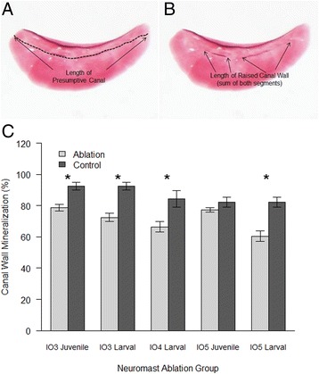
Figure 5.

Percentage of canal wall mineralization following infraorbital canal neuromast ablation in adults. (A) Diagram showing the total length of canal (dashed line) and (B) length of mineralized canal wall (arrows) in an adult infraorbital three. (C) Bar graph summarizing the percent of the IO canal length that formed a mineralized wall (y-axis). Error bars show standard errors. Significance is indicated by asterisk where ablation groups differed from controls (no ablation): p < 0.05 in larval specimens for IO3 (t(23) = 0.329), IO4 (t(22) = 4.19) and IO5 (t(21) = 4.69) bones. Juvenile IO3 ablation specimens were also significantly different (t(18) = 4.215, p < 0.05).
