
Figure 7.
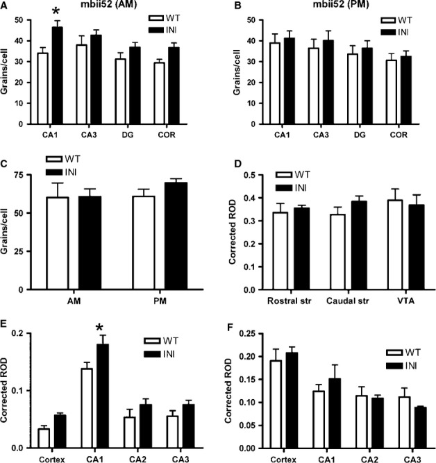
In situ hybridisation revealed compensatory gene expression changes in INI mouse brain. mRNA levels were measured by in situ hybridisation in INI and WT mice (n = 5–8) and silver grains per cell were quantified in brain regions (A-C) in the morning and evening, or signal intensity was quantified by film densitometry (D–F) in the morning samples only. Expression of mbii52 snRNA was increased in INI mice in the morning (A) but not in the evening (B). The brain-specific tryptophan hydroxylase 2 (TPH2), in raphe nuclei, was not altered by the lack of editing (C). The dopamine receptor 2 levels were similar in all regions tested (D). Two other 5-HT receptors were quantified; 5-HT1A levels were higher in INI mice (E) but 5-HT2A levels were not (F). All data were analysed by two-way anova, values are mean + SEM; *P < 0.05. CA, hippocampal cornu ammonis; COR, cortex; DG, hippocampal dentate gyrus; ROD, relative optical density; str, striatum; VTA, ventral tegmental area.