
Figure 2.
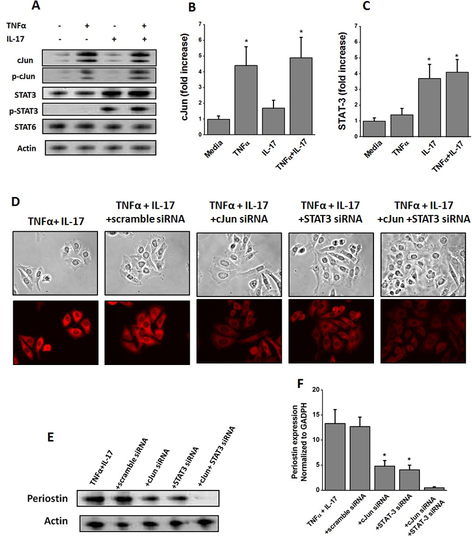
Induction of transcription factors cJun and STAT-3 in HepG2 cells following stimulation with TNFα and IL-17, respectively. (A) Western blot analysis was performed to determine the protein level expression of cJun (43 kDa), p-cJun (43 kDa), STAT-3 (86 kDa), p-STAT-3 (86 kDa), STAT-6 (116 kDa), and Actin (43 kDa) for both total and active phosphorylated forms analyzed after 30 min stimulation with TNFα, or IL-17 or both; (B and C) quantitative mRNA expression of cJun (B), and STAT-3 (C) by qRT-PCR analyzed after 30 min stimulation with TNFα, or IL-17 or both. Quantitation done by ΔΔcT method normalized for GADPH expression. (D) Immunostaining of HepG2 cells with periostin following treatment with with both TNFα and IL-17, and specific siRNA knock down of cJun and STAT-3. (E) Western blot analysis of perisotin expression in the hepG2 cells following stimulation with TNFα and IL-17 along with cJun and STAT-3 knock-down by specific siRNA. Scramble siRNA usd as negative control. (F) Periostin mRNA expression analyzed by quantitative RT-PCR in the hepG2 cells following stimulation with TNFα and IL-17 along with cJun and STAT-3 knock-down by specific siRNA. Quantitation done by ΔΔcT method normalized for GADPH expression. All data represented as mean values ± SEM from four independent experiments.