
Figure 4.
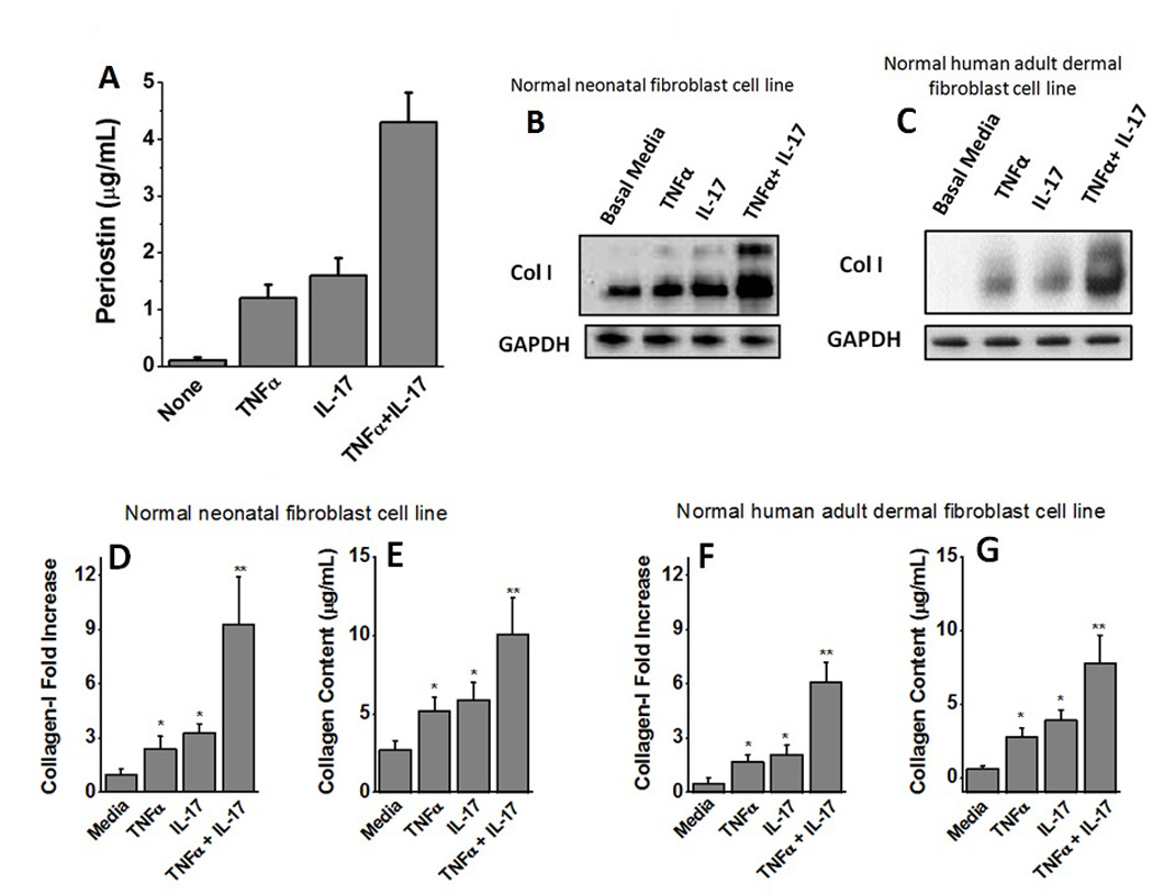
Induction of Collagen expression following activation of fibroblasts with periostin rich supernatant. (A) Quantitative ELISA analysis of the periostin secreted into the supernatant by HepG2 cells following stimulation with TNFα, or IL-17, or both. (B) Western blot analysis of the Type-I Collagen expressed by the normal neonatal human fibroblasts following treatment with supernatant from HepG2 cells stimulated with TNFα, or IL-17 or both. (C) Western blot analysis of the Type-I Collagen expressed by the normal adult human dermal fibroblasts following treatment with supernatant from HepG2 cells stimulated with TNFα, or IL-17 or both. (D) Densitometric analysis to determine quantitative expression of Collagen-I in normal neonatal human fibroblasts (under various conditions mentioned in B). (E) Sircol assay to demonstrate the expression of soluble collagen by fibroblasts in normal neonatal human fibroblasts (under various conditions mentioned in B). (F) Densitometric analysis to determine quantitative expression of Collagen-I in normal adult human dermal fibroblasts (under various conditions mentioned in C). (G) Sircol assay to demonstrate the expression of soluble collagen by fibroblasts in normal adult human dermal fibroblasts (under various conditions mentioned in C). All data represented as mean values ± SEM from four independent experiments.