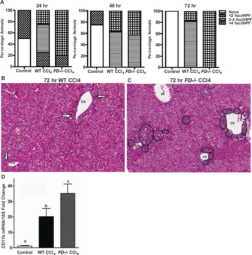
Figure 5. FD−/− mice have higher presence of hepatic neutrophilic infiltrates following CCl4- induced injury.
A. Identification of neutrophilic infiltrates evaluated from histological Hematoxylin and Eosin (H&E) sections of livers from WT and FD−/− mice at 24, 48 and 72 hr following a single injection of CCl4 are shown. Bars represent percentage of mice in each group exhibiting the number of foci observed per high power field. The number of mice at each time point are the same as those indicated in Figure 3. B &C. Representative histological H&E sections of livers from WT and FD−/− mice exhibiting the most frequent number of neutrophilic foci at 48 hr after a single injection of CCl4 are shown (H&E stain, 200×) (CV: central vein; PT: portal tract). B. Arrows indicate single foci with scattered neutrophils and rare associated lymphocytes noted in the centrilobular region in WT mice. C. Circles indicate the numerous zone 3 clusters of neutrophils seen in the FD−/− mice D. mRNA expression of CD11b was detected in mouse liver 48 hr after a single injection of CCl4. Values represent means ± SEM. Values with different alphabetical superscripts were significantly different from each other, p<.05.

