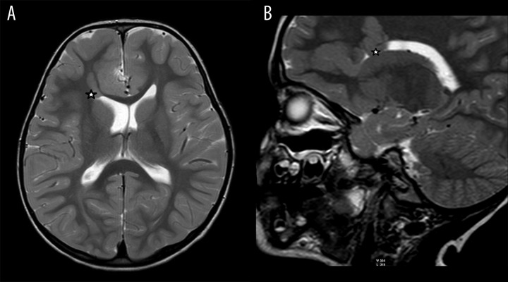
Figure 2.
A 5-year-old boy. Clinical symptoms: retarded speech development. MR image. (A) T2-weighted image, axial plane. (B) T2-weighted image, sagittal plane. Heterotopic gray matter within the white matter of the right cerebral hemisphere and in the subependymal region of the frontal horn of the right lateral ventricle (asterisk).

