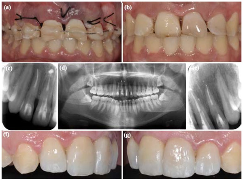
Figure 3.
(a) Surgical repositioning and semi-rigid splinting using orthodontic wire-composite. (b) Splint was removed. (c) Root canal treatment and filling with glass fiber posts in 1.1 and 1.2. (d) Orthopantomography, 2 weeks follow-up. (e) Root canal treatment and filling with fiberglass posts in 2.1 and 2.2. (f) Lateral left view, the traumatized teeth were restored with minimally invasive preparations of feldspathic ceramic. (g) Lateral right view.

