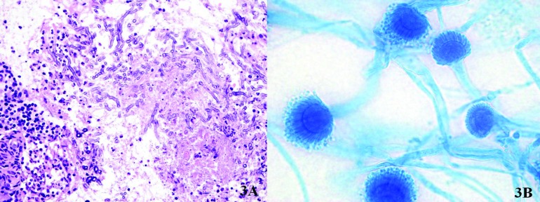
Figure 3.

A.Histological examination (hematoxylin and eosin stain) shows abundant septate fungal hyphae with dichotomous branching suggestive of the Aspergillus spp., with non-caseating granulomatous inflammation, eosinophils and giant cells; B. Microscopic morphology of A. fumigatus (100x; lactophenol blue stain): hyphae are septate and the coniodophore is enlarged at the tip, forming a swollen vesicle.
