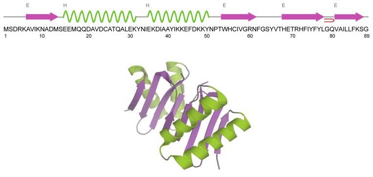
Figure 1.

Amino acid sequence, secondary structure (top) and 3D X-ray structure (bottom) of Drosophila dynein light chain LC8. The structure is generated from PDB entry file 3DVT (Lightcap et al. 2008). LC8 is shown as homodimer. The α-helices are shown in green and β-sheets are in purple.
