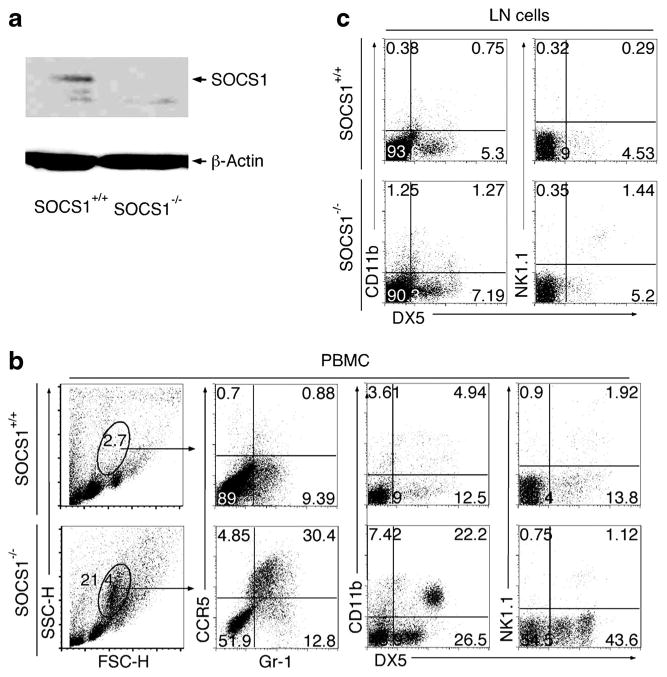
Fig. 1.

Characterization of innate immune cells of SOCS1-KO mice. a Western blot analysis of protein extracts from PBMC of control or SOCS1KO mice. Freshly isolated PBMC (b) and LN (c) cells from control or SOCS1KO mice were stained with labeled monoclonal Abs specific to cell surface markers of innate immune cells and analyzed by FACS. Numbers in quadrants represent percentage of cells expressing cell surface markers indicated on figure. Data represent at least three independent experiments.
