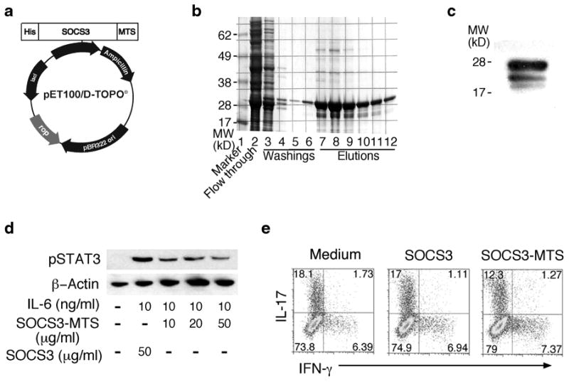
Fig. 6.

Cell-penetrating SOCS3 (MTS-SOCS3) suppressed the expansion of Th17 cells. a Schematic illustration of MTS-SOCS3 expression vector. b Coomassie blue stained gel at various stages of purification of the MTS-SOCS3.c Western blot analysis of purified MTS-SOCS3. d Macrophages were loaded with MTS-SOCS3 or GST-SOCS3, stimulated with IL-6 for 15 min, and analyzed by Western blotting. e Th17 uveitogenic T cells were cultured in medium containing MTS-SOCS3 or SOCS3 for 4 days, and IL-17 or IFN-γ expression was detected by intracellular cytokine staining and flow cytometry. Data represent three independent experiments.
