
FIGURE 2.
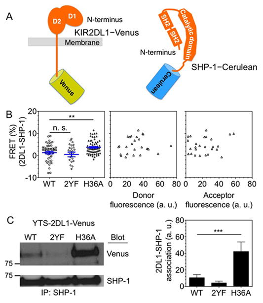
H36A mutation of KIR2DL1 results in constitutive SHP-1 recruitment. (A) Diagram of KIR2DL1 and SHP-1 tagged at their C-terminus with Venus and Cerulean, respectively. (B) SHP-1 proximity to KIR2DL1 monitored by FRET. SHP-1 Cerulean was transiently expressed in YTS cells stably transfected with 2DL1-WT-Venus, 2DL1-2YF-Venus, or 2DL1-H36A-Venus. FRET signal at the plasma membrane was calculated as percentage increase in donor (Cerulean) fluorescence after acceptor (Venus) photobleaching. The negative values of FRET signal are due to unintentional photobleaching of donor fluorescence during acquisition. In the left-most panel, data points from three independent experiments are combined. Horizontal and vertical lines represent the mean and standard error, respectively. The middle and right-most panels show FRET signals of SHP-1 with 2DL1-H36A relative to the expression of FRET donor (SHP-1-Cerulean) and FRET acceptor (2DL1-H36A-Venus). (C) Endogenous SHP-1 was immuno-precipitated from the indicated YTS-2DL1-Venus cells and its association with 2DL1 was probed with an anti-GFP immunoblot. The right panel shows band intensities obtained by scanning immunoblots and calculated using ImageJ. The error bars represent spread in the values of mean obtained from two independent experiments.