
FIGURE 6.
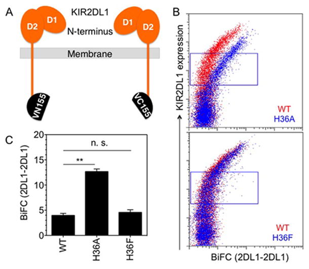
A bulky side chain at amino acid 36 of KIR2DL1 prevents constitutive receptor self-association. (A) Diagram of KIR2DL1 tagged at the C-terminus with Venus fragments VN155 and VC155. (B) Constitutive KIR2DL1 self-association in transfected YTS cells monitored by BiFC. BiFC signal of 2DL1-WT (red) and 2DL1-H36A (top) or 2DL1-H36F (bottom) (blue) is plotted against surface expression, as detected by anti-2DL1-APC binding. (C) Median fluorescence intensity of BiFC signal in the gates shown in panel B. The error bars represent spread in the values of median BiFC signal determined from two independent experiments.