
Figure 3.
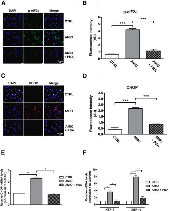
AMIO induces UPR activation in ML-1 cells. A and C, Patterns of expression of phospho-eIF2α and CHOP in ML-1 cells as determined by immunostaining. ML-1 cells were grown on glass coverslips for 24 hours and then pretreated or not with 7.5 mM PBA for 24 hours followed by 24 hours in medium with 5 μM AMIO. Immunofluorescence staining was performed using phospho-eIF2α antibody (green) or CHOP antibody (red). Nuclei were counterstained with DAPI (blue). B and D, Quantification of fluorescence intensity for phospho-eIF2α or CHOP in cultured ML-1 cells treated as described. E and F, The levels of mRNAs for CHOP, XBP-1, and active XBP-1 spliced mRNA were determined by real-time RT-PCR analysis of total RNA from ML-1 cells treated as above. GAPDH was used as an internal standard. Each bar represents the mean ± SEM of four independent experiments, each performed in triplicate. *, P < .05; **, P < .01, ***, P < .001 compared with vehicle-treated cells (CTRL).