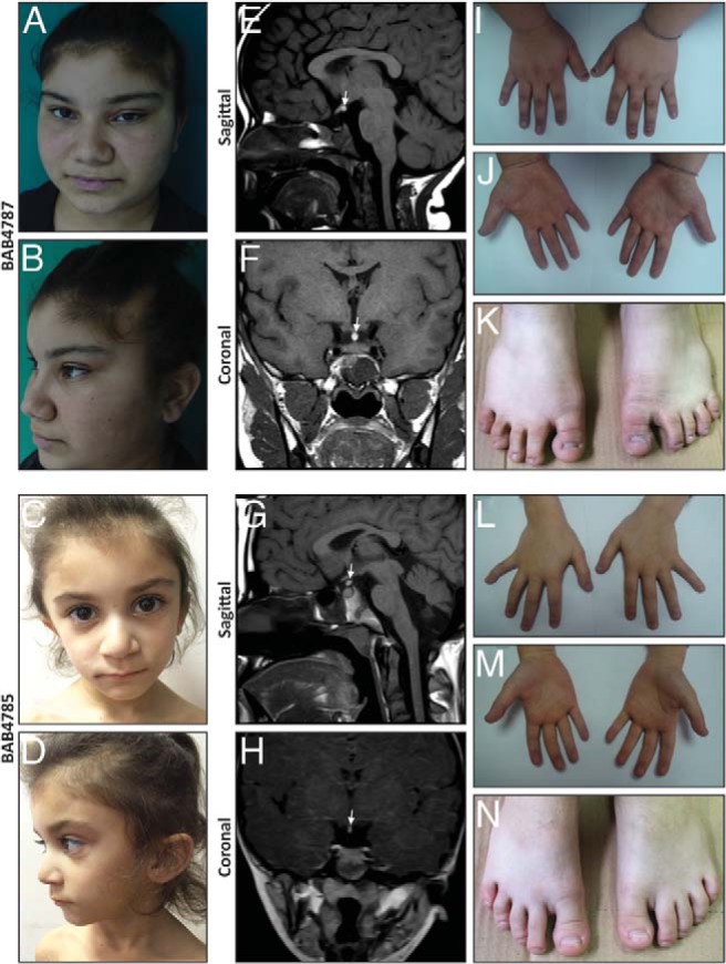
Figure 1.

Facial images, extremity pictures, and hypophysis MRIs of the patients. A–D, Pictures of both affected siblings show hypotelorism, sparse hair on the frontal region, broad nasal root, and thick ala nasi. E–H, Hypophysis MRI reveals thin pituitary gland together with ectopic neurohypophysis and interrupted stalk. I–N, Pictures of hands and feet show partial syndactyly of second and third toes and hypoplastic nails.
