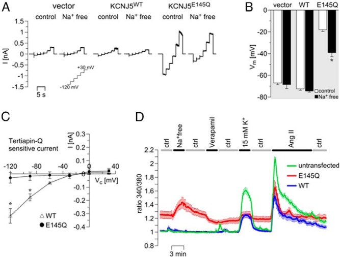
Figure 2.
The Kir3.4 p.Glu145Gln mutation induced a Na+ influx and increased intracellular Ca2+ levels in adrenal NCI-H295R cells. Representative whole-cell currents, A; and membrane voltage, B, of NCI-H295R cells transfected with empty vector (n = 5), WT KCNJ5 (WT, n = 10), or mutant KCNJ5 encoding the p.Glu145Gln substitution (E145Q, n = 16). Currents and voltage were measured under control (ctrl, normal Ringer solution) and Na+-free (using N-methyl-D-glucamine chloride, NMDG+, instead of NaCl) conditions. C, The tertiapin-Q (1μM) sensitive current of cells expressing the KCNJ5 WT (WT; n = 12) or mutant KCNJ5 encoding the p.Glu145Gln substitution (E145Q; n = 6) is shown. To stimulate currents in cells expressing the WT channel, the pipet solution was supplemented with Na+ (30mM) and GTP (0.5mM) and bath K+ was increased (50mM K+). D, Fura-2 Ca2+ measurements: Traces show mean values of 340 nm/380 nm ratios ± SEM as a measure of intracellular Ca2+ concentration (WT in black, E145Q in red, and untransfected cells in green; n = 17–26 per group) under control and Na+-free conditions. Verapamil (10μM) and angiotensin II (Ang II, 20nM) were applied in control solution. In the solution containing 15mM K+, the Na+ concentration was reduced to the same extent. *, P < .05 comparing B, control and Na+-free; or D, WT and Kir3.4- p.Glu145Gln-expressing cells.

