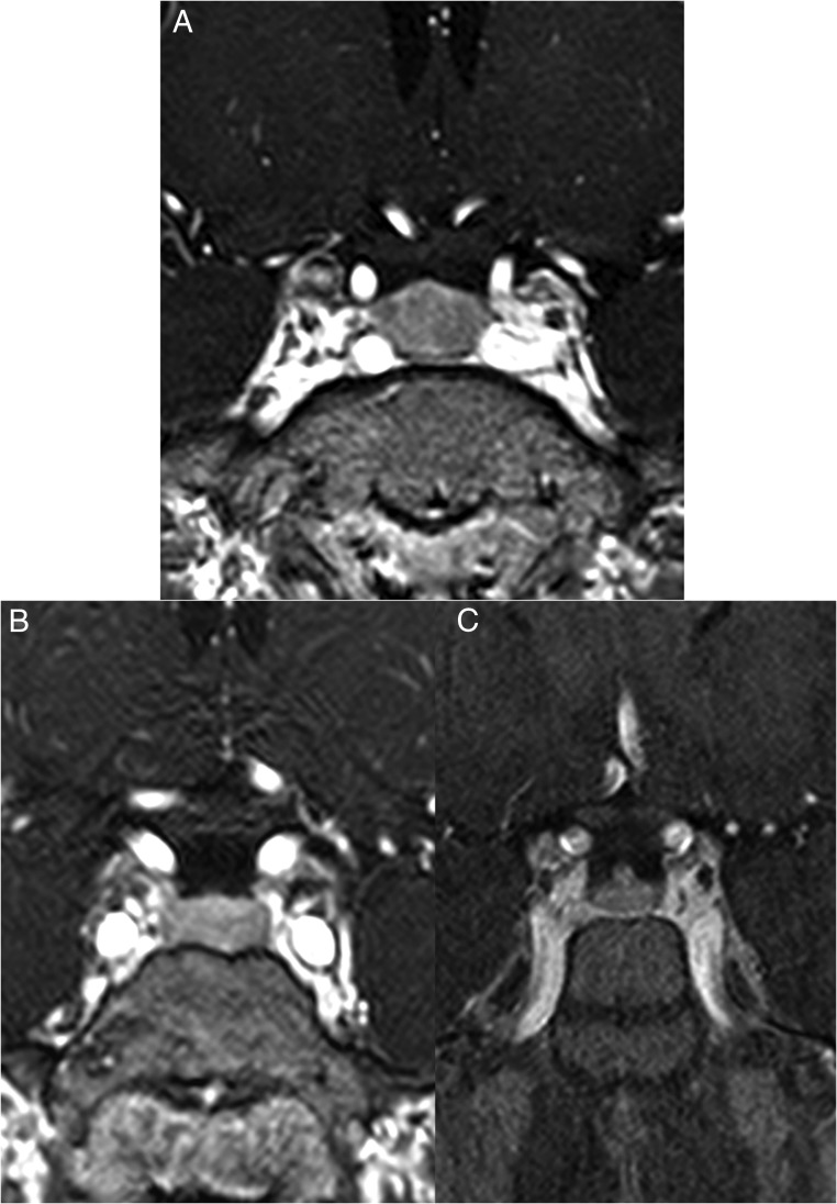
Figure 3.
A, Coronal post contrast T1-weighted 3D gradient echo MRI scan of patient 3. The pituitary gland is slightly enlarged with the vertical height measuring 10.5 mm. There is homogeneous enhancement throughout the pituitary parenchyma. These findings suggested diffuse hypertrophy. No focal space occupying lesion was present within the gland; (B, C) Two coronal post contrast T1-weighted 3D gradient echo MRI scans performed before (B) and after (C) surgical resection of the CRH/ACTH-secreting tumor of patient 2. After resection of the tumor, there is obvious reduction in the size of the pituitary (C).

