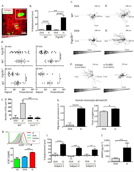
Figure 2. Immune complex FcγR engagement promotes chemokine-directed mouse and human DC migration in vitro.
(a) Representative image of WT bone marrow-derived DCs ((BMDCs), GFP, green) embedded in a collagen matrix (red) prior to the application of a CCL19 gradient. (b) X-displacement of WT and Fcgr2b−/− BMDCs incubated with ovalbumin (OVA) or IgG-opsonised ovalbumin (IC) for 24 hours prior to incorporation within a collagen matrix and subsequent exposure to a CCL19 gradient. Graphs show mean and SEM of values obtained from one collagen gel, representative of at least three experimental replicates. (c) Representative migration tracks in a CCL19 gradient of WT (upper panels) and Fcgr2b−/− (lower panels) BMDCs incubated with OVA (left panels) or IC (right panels) for 24 hours prior to suspension in the collagen matrix. (d) Angle of leading protrusion relative to chemokine gradient of WT (open circles) and Fcgr2b−/− (filled squares) BMDCs suspended in a collagen matrix at 10, 20, 40, and 60 minutes following application of CCL19 gradient. Angles closer to 0° indicate cell polarization parallel to the gradient. (e) Representative migration tracks in a CCL19 gradient of BMDCs incubated with IC, either in the presence of an FcγRIII blocking antibody (right panel) or an isotype control antibody (left panel). (f) Quantification of number of WT and Ccr7−/− DCs within draining lymph nodes 48 hours following subcutaneous transfer. Mean and SEM from 6 lymph nodes from 3 mice in each group shown. (g) CCR7 expression on human monocyte-derived DCs incubated with OVA (blue line) or IC (red line) for 24 hours. Representative histograms are shown in the upper panel and GMF and SEM of triplicates from one of three replicates shown in the lower panel. (h) Migratory persistence in X (left panel) and track straightness (right panel) of human monocyte-derived DCs in a human CCL19 gradient following incubated with OVA or IC for 24 hours prior. Values represent the mean and SEM from cells migrating in at least three collagen gels per subject and from three healthy subjects. (i) X-displacement of human monocyte-derived DCs from 3 subjects following application of a CCL19 gradient. Values represent the mean and SEM. (j) MMP-9 levels in culture supernatants obtained from human monocyte-derived DCs isolated from 3 healthy subjects. Values represent the mean and SEM. In (c) and (e), tracks are derived from a single collagen gel and representative of three similar experiments. P values calculated using a Students t test (two-tailed). * = P ≤ 0.05, ** = P ≤ 0.01, *** = P ≤ 0.001, **** = P ≤ 0.0001.

