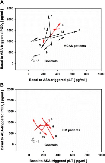
Figure 3.

Vector plots of ASA-triggered PGD 2 and pLT dynamics derived from PBLs of MCAS and SM patients and healthy controls. Origin of arrows: basal eicosanoid release; arrowheads: ASA-triggered eicosanoid release; grey arrows: healthy individuals (n = 20); black arrows: MCAD-M, MCAD patients without MCAD-specific medication (n = 9); red arrows: MCAD + M, MCAD patients with MCAD-specific medication (n = 12); black numbers: patients’ identification numbers (see Table 1). (A) MCAS: MCAD patients with mast cell activation syndrome (n = 12); (B) SM: MCAD patients with systemic mastocytosis (n = 9). One SM patient was not depicted in the figure, because of extraordinarily high pLT release, both basal and after ASA exposure (basal PGD2 and pLT release: 531 and 258 pg/ml, respectively; ASA-triggered PGD2 and pLT release: 2056 and 853 pg/ml, respectively).
