Figure 1.

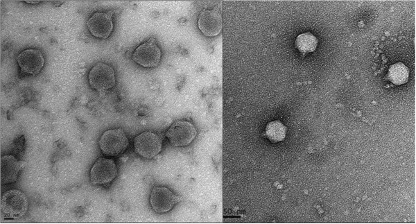
Electron micrographs of phage AP5. The phage has been negatively stained with 2% potassium phosphotungstate. AP5 is shown at 50,000× magnification or 150,000× magnification. Scale bar indicates size in nm.

Electron micrographs of phage AP5. The phage has been negatively stained with 2% potassium phosphotungstate. AP5 is shown at 50,000× magnification or 150,000× magnification. Scale bar indicates size in nm.