Figure 2.
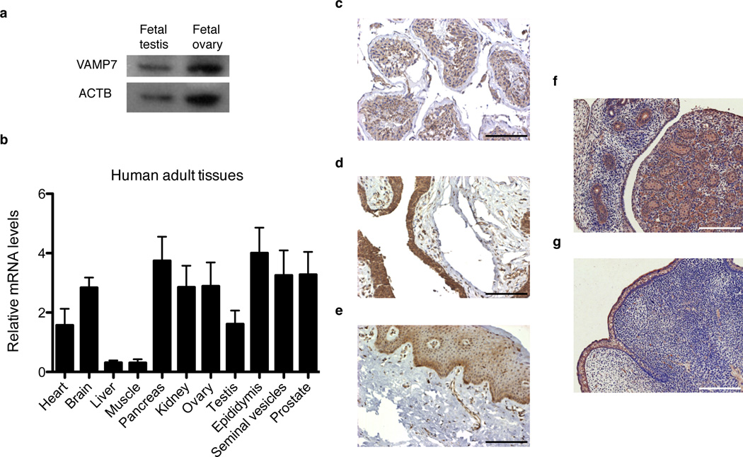
VAMP7 is present in human and murine male reproductive tissues. (a) Western-blot analysis of VAMP7 expression in human fetal testes and whole ovarian lysates. ACTB (β actin) was used a loading control. (b) qRT-PCR analysis of VAMP7 mRNA levels normalized to GAPDH in human adult tissues. n = 6 independent samples for each tissue. Data are presented as means ± s.e.m. (c–e) Immunohistological detection of VAMP7 in normal human adult testicular (c) and penile tissues including urethral (d) and preputial (e) epithelia. Scale bar, 125 µm. (f,g) Immunohistochemical detection of VAMP7 expression in fetal testis (f) and external genitalia (g) from wild-type male mice at embryonic stage E16.5. Scale bar, 125 µm.

