Figure 1.
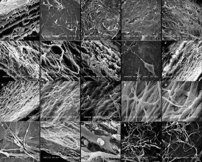
Cell morphology and the network formed (scanning electron microscopy).
BMSCs were grown on FBADM either non-induced in basal medium for 12–34 days (spontaneous differentiation) or induced in basal medium for 3 days and then neural differentiation medium for 9 days, basal medium for 3 days and then for 31 days, or basal medium for 17 days and then neural differentiation medium for 17 days. (A–C) The structure of the FBADM. (A) A longitudinal section. (B) The surface of the FBADM with an intact basement membrane. (C) The surface of the FBADM with a damaged basement membrane. (D) Of the non-induced cells cultured in basal medium for 12 days on FBADM, most grew parallel to the flat surface. (I) The cells induced in basal medium for 3 days and then neural differentiation medium for 9 days on FBADM, showing the long processes of typical neurons, an uplifted cell body, and unipolarity. (E–H) The non-induced cells cultured in basal medium for 34 days on FBADM showing proliferation, spontaneous differentiation, and stratified growth (E); neuronal-like differentiation (F); vascular-like differentiation (G); and matrix morphology changes (H) compared with the structure of the original (A). (J–O) The BMSCs were induced in basal medium for 3 days and then neural differentiation medium for 31 days on FBADM, showing (J) the stratified growth of the differentiated cells with FBADM at lower magnification; (K) the differentiated cells connected to each other and into the mesh; (L, M) the differentiated cells formed a complete neural circuit with the formation of a growth cone and synaptic connections by dendrites and axons; and (N–O) nerve fibers formed by a parallel arrangement of microfibers at high magnification. (P–T) When the BMSCs were induced in basal medium for 17 days and then neural differentiation medium for 17 days on FBADM, the differentiated cells formed a crisscross neural network-like structure on the surface of the scaffold (P) and inside (longitudinal section, Q) a network connection. (R) A higher magnification image of Q displaying the synaptic connections and dendritic spines. (S, T) The long smooth axons, growth cone, and axon-dendritic and axon-body synaptic connections of the neural network-like structure. BMSCs: Bone marrow mesenchymal stem cells; FBADM: fetal bovine acellular dermal matrix.

