
Figure 1.
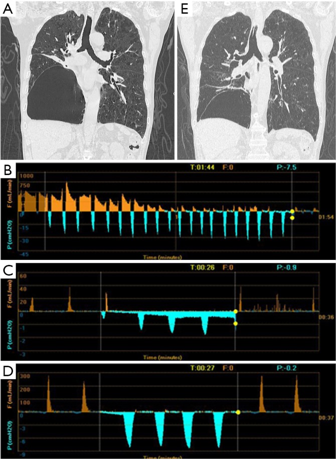
Chest CT and Chartis result of patient 1. (A) A bulla in the RLL and atelectasis of right RML; (B) drop of airflow to zero indicated CV is negative at RUL; (C) Chartis test showed low flow at RML; (D) Chartis test showed low flow at RLL; (E) the volume of bulla reduced and the atelectasis of RML disappeared. RLL, right lower lobe; RML, right middle lobe; CV, collateral ventilation; RUL, right upper lobe.