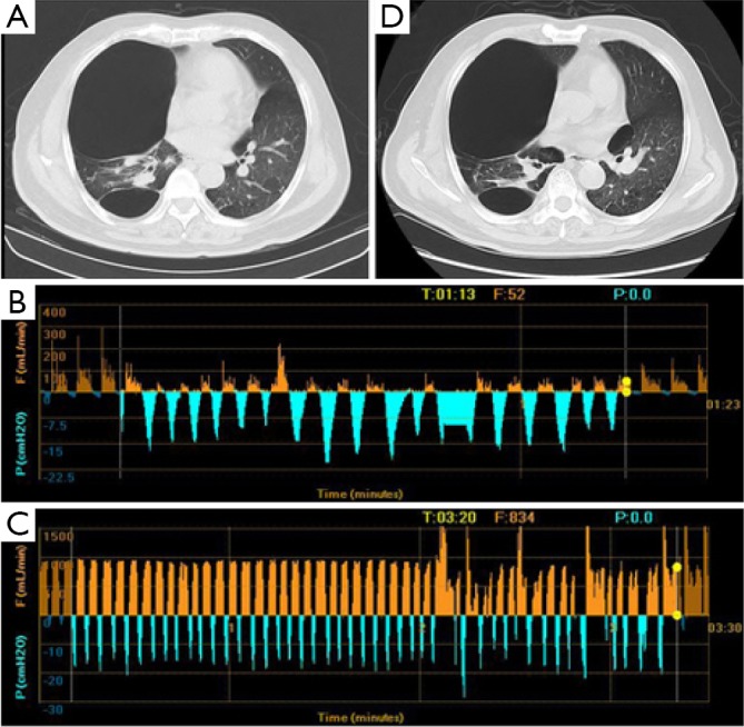
Figure 2.

CT imaging and Chartis result of patient 2. (A) A bulla in RUL and severe destruction of lung tissue of RUL; (B) Chartis test showed low flow at RUL; (C) CV was positive as measured by Chartis system at RLL; (D) chest CT did not show reduction of bulla volume at RUL. RUL, right upper lobe; CV, collateral ventilation; RLL, right lower lobe.
