
Figure 5.
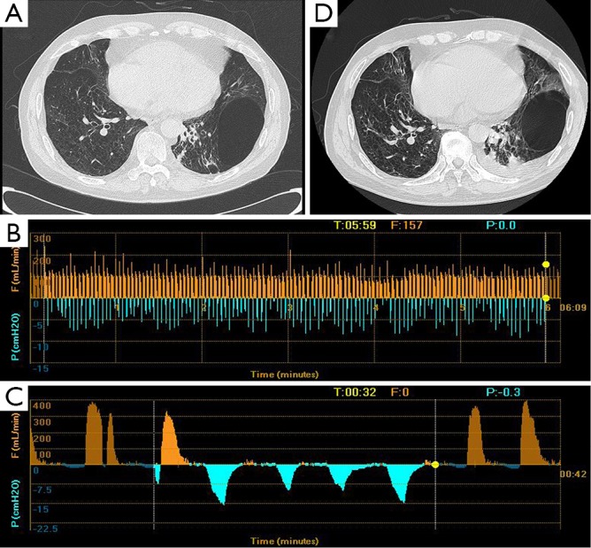
Clinical features of patient 5. (A) CT scan showed a bulla at the position of LLL; (B) the Chartis assessment showed the presence of CV at LUL; (C) low flow at LLL; (D) the volume of the bulla did not change after the EBV placement. CV, collateral ventilation; LLL, left lower lobe; LUL, left upper lobe; EBV, endobronchial valve.