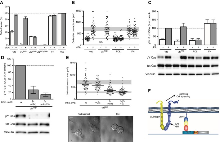
Figure 2. The RGD motif VN is required for integrin-mediated cell adhesion but dispensable for uPAR-induced β1 integrin signalling and cell spreading.
A Effect of VN variants on uPAR and integrin-dependent cell adhesion. 293 uPART54A cells were seeded, in the presence or absence of uPA, on wells coated with WT VN or recombinant VN variants in which the integrin binding site is inactivated (VNRAD), the SMB domain is deleted (VNΔSMB) or both (VNRADΔSMB) for 30 min. Cell adhesion was assayed and expressed as percentage of PDL. For comparison, the adhesion to fibronectin (FN) and PDL is shown. The data are means ± s.e.m. (n = 3).
B, C uPAR-induced cell spreading and signalling do not require vitronectin integrins engagement. 293/uPART54A cells were seeded on the indicated substrate with or without uPA for 30 min. Cell-matrix contact area (B) and p130Cas SD phosphorylation (C) were analysed and quantified. Cell-matrix contact area measurements represent at least 50 cells with indicated geometrical means ± 95% CI, in two independent experiments. Cells (n = 10) having contact areas exceeding the scale of the y-axis are not shown. Cells do not adhere to VNRAD in the absence of uPA, and the quantification of cells spreading is therefore not applicable (n.a.). The grey bars represent the range of cell area of not spread or fully spread uPART54A cells based on 95% CI on poly-D-lysine or on VN with uPA, respectively. Western blot data are represented as percentage of phosphorylated p130Cas SD of cells treated with uPA on VN (n ≥ 6, mean ± s.e.m.). Representative blots are shown.
D, E β1 integrin mediates uPAR-induced signalling and spreading on an integrin refractory VN variant. 293/uPART54A cells were treated with the indicated antibody and seeded on VNRAD with uPA for 30 min. p130Cas SD phosphorylation (D) and cell-matrix contact area (E) were analysed and quantified. Cell-matrix contact area measurements represent at least 50 cells with indicated geometrical means ± 95% CI, in two independent experiments. The grey bars represent the range of cell area of not spread or fully spread uPART54A cells based on 95% CI on poly-D-lysine or on VN with uPA, respectively (B). Representative DIC images are shown (scale bar: 10 μm). The Western blot data are represented as percentage of phosphorylated p130Cas SD of cells seeded on VNRAD with uPA in absence of antibody (n ≥ 3, mean ± s.e.m.). Representative blots are shown.
F Cartoon illustrating the β1 integrin signalling triggered by uPAR on VNRAD.

