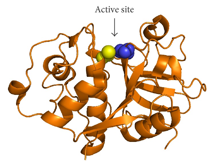
Figure 1.

The papain-like peptidase fold illustrated on the crystal structure of papain. The protein is shown in cartoon representation and the position of the active site cleft is marked by an arrow. Catalytic residues Cys and His are shown as yellow and blue spheres, respectively. Coordinates were obtained from the Protein Data Bank under accession code 1PPN. The image was created with PyMOL (Schrödinger, LLC, Portland, OR, USA).
